
In biological tissue, electric current affects the components and structures, which have an electric charge and/or electric dipole moment. In the space surrounding cell membranes from both sides, it is the electric charges presented mostly by ions and sources of dipole moment presented by polar molecules of water and mobile polar macromolecules act as the sources of changes. It is the so-called extramembraneous electric conductivity. Active resistance in such case of the biological object depends on the chemical structure of this object, to be more exact, on the chemical compounds filling the extramembraneous space. The active resistance component is in small dependence of the frequency, it may be considered constant in the frequency range 10 Hz – 10 MHz, it is the so-called frequency-independent component.
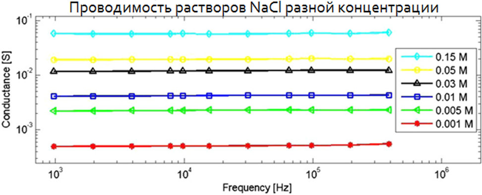
Ionic solutions such as aqeous solution of NaCl do not demonstrate dielectric dispersion at frequencies below 1 MHz.
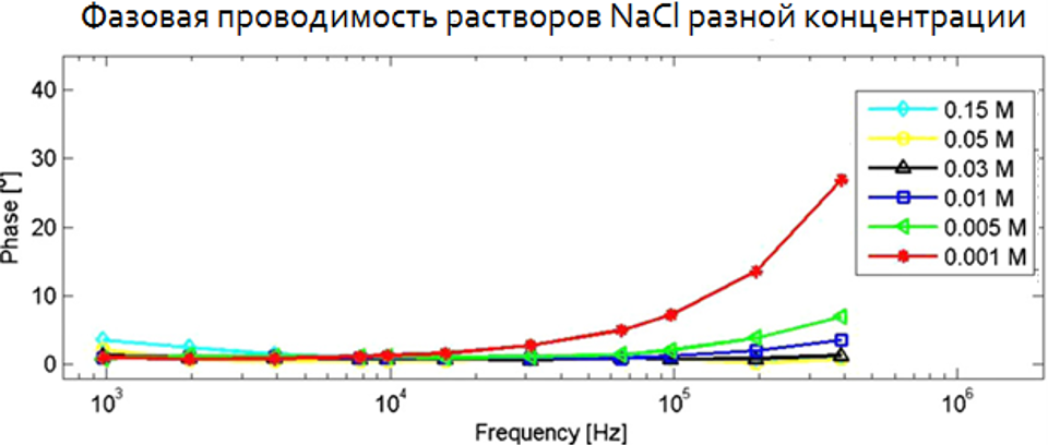
Ionic solutions demonstrated dielectric dispersion only at frequencies above 1 MHz.
The ability of the cellular membrane for polarization due to protein and lipidic structures determines its exceptional electrical properties. Membrane electrical conductivity associated with the double lipidic layer is measured through the reactive resistance of the biological object. The capacitive reactance reveals strong dependency of frequency, it is the so-called frequency-dependent component. At frequencies below 100 kHz the capacitive reactance s 2 or 3 times less than the active component of impedance.
Therefore, saline solution or other saline solutions are purely resistive materials, i.e. frequency-independent, and, consequently, multi-frequency electrical impedance systems cannot examine saline solutions in a proper manner because the response of purely resistive materials does not change along with frequency change. And protein-lipidic structure of biological cell membrane creates capacitance of the cell and allows to apply multi-frequency electrical impedance system for assessment of the biological object using current frequency in the range of 100-500 kHz.
The method of medical diagnostics which calculates electrical conductivity or dielectric permittivity of a body part based on superficial electrical measurements is called electrical impedance spectroscopy. Electrical impedance is the term used to designate resistance of an object to electrical current, which has frequency-dependent and frequency-independent properties.
Some cancer cells demonstrate frequency-dependent change of impedance, which differs significantly from the impedance of healthy cells. That is the reason why measurement of electrical impedance at different frequencies can be used for diagnostics of tumours.
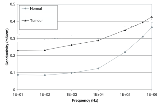
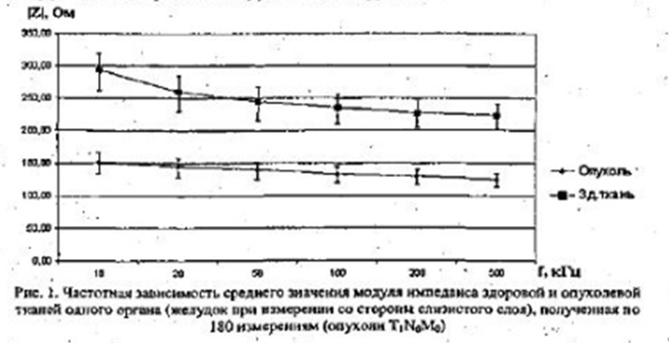
The graph above shows that average conductivity of tumour tissue is significantly higher than that of healthy tissue throughout the whole range of frequencies (from 10 Hz to 1 MHz).
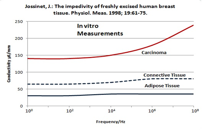
The graph shows that during in vitro studies breast carcinoma demonstrates electrical conductivity three times higher than that of connective and adipose tissues. With the increase of frequency this ratio practically does not change (Jossinet, J.: “The Impedivity of Freshly Excised Human Breast Tissue”, Physiol. Meas. 1998; 19:61-75).
The graph shows that the tumour impedance was detected in the study to be significantly lower than that of healthy tissues, and with the increase of frequency this ratio practically did not change (“Methods and Means of Multi-Frequency Electric Impedancometry of Human Tissue for Cancer Surgery” K.D. Belik 2010).
The distinguishing feature of impedance spectroscopy is the use of wideband equipment (from 10 kHz to 1 GHz).
MEIK electrical impedance mammograph is a tomographic visualization system and uses the following parameters of electric current: current 0.5 mA, frequency 50 kHz. Such parameters allow to receive a good-quality image with good resolution and qualitative information. During testing of the system in the Institute of Radiotechnology and Electronics of the Russian Academy of Science and after clinical studies held by SIM-Technika specialists the work frequency of 50 kHz, optimal from the point of view of convenience of use and the level of parasitic signals, was chosen. These tests showed that the quality of visualization (resolution, sensitivity) remain stable for work frequencies 50 kHz. At higher frequencies the effect of artifacts becomes noticeable, at frequencies below the work frequency it becomes more complicated to solve the issue of high impedance at the electrode-skin transfer.
Application of electrical impedance mammograph with current force of 0.5 mA and frequency of 50 kHz allows to assess extramembrane frequency-independent electrical conductivity. Other things being equal (mostly external factors, i.e. current force, current frequency), the level of electrical conductivity and the electrical impedance image itself depend on the extramembrane concentration of ions and their travel rate.
Application of MEIK single-frequency electrical impedance mammograph has demonstrated its efficiency for early diagnostics of breast cancer.
Sachin Prasad N, Houserkova D, Campbell J. Breast imaging using 3D electrical impedence tomography. Biomedical Papers of the Medical Faculty of the University Palacky, Olomouc, Czech Republic. 2008;152(1):151-154
Raneta O, Bella V, Bellova L, Zamecnikova E. The use of electrical impedance tomography to the differential diagnosis of pathological mammographic/sonographic findings. Neoplasma. 2013;60(6):647-654
Daglar G, Senol K, Yakut ZI, Yuksek YN, Tutuncu T, Tez M, et al. Effectiveness of breast electrical impedance imaging for clinically suspicious breast lesions. Bratislava
Medical Journal. 2016;117(9):505-510
Feng X, Mengxin L, Peter J, Hongchuan J. Utilisation of electrical impedance tomography and/or ultrasound and mammography in breast disease diagnosis: A controlled study. National Medical Journal of China. 2017;97(18):1391-1395
Murillo-Ortiz B, et al. Diagnosis of breast cancer by electrical impedance mammography MEIK. Revista Mexicana de Mastologнa. 2019;9:20-27
Alexander Karpov, Marina Korotkova, Gregory Shiferson and Elena Kotomina.
Electrical Impedance Mammography: Screening and Basic Principles. “Breast Cancer and Breast Reconstruction” Edited by Luis Tejedor. 2020
Electrical impedance mammography should be a method of primary breast cancer screening due to its high informativeness, safety for the personnel and the patient, its portability and mobility.
Electrical impedance mammography is a means of formation of breast cancer survey or risk groups for the purpose of dynamic monitoring. It is possible to organize risk groups due to the possibility to perform quantification of the condition of the mammary gland.
Electrical impedance mammography performs its functional screening tasks without ionizing radiation and other potentially harmful methods.
It can be used for examining women of all age groups in policlinics and outpatient clinics, schools, women’ care clinics, maternity hospitals, sanatoriums, i.e. in places with a lot of women there.
Therefore, the problem of organization of large-scale screenings for women can be easily solved.
